Muscles Of The Chest And Abdomen Labeled : Abs Muscle Anatomy Abdomen Muscle Anatomy - Human Anatomy ... / Pronounced muscular and chest pain.
byAdmin•
0
Muscles Of The Chest And Abdomen Labeled : Abs Muscle Anatomy Abdomen Muscle Anatomy - Human Anatomy ... / Pronounced muscular and chest pain.. Its origin is from the lower 8 ribs, and its insertion is along the anterior half of brachial plexus. Their main function is contractibility. The abdomen (colloquially called the belly, tummy, midriff or stomach) is the part of the body between the thorax (chest) and pelvis, in humans and in other vertebrates. This requires complete exposure of the region in question. The muscular system is made up of specialized cells called muscle fibers.
Some of the signs and symptoms include: The upper part of the trunk is the chest and the lower one is the abdomen. The primary function is certainly to provide support to the skeletal system and to facilitate its movements. There are three muscular layers of the abdominal wall, with a fourth layer in the middle anterior region. Small muscles running between the ribs, known as the external intercostal muscles, lift the ribs during deep breathing to further expand the chest and lungs and provide even more air to the body.
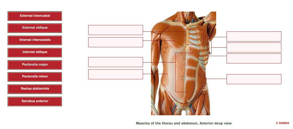
anatomy human abdomen | MRI abdomen coronal anatomy | free ... from i.pinimg.com The external oblique muscle is a broad muscle that runs along the anterolateral abdomen and chest wall. The abdomen (colloquially called the belly, tummy, midriff or stomach) is the part of the body between the thorax (chest) and pelvis, in humans and in other vertebrates. They are the pectoralis major, pectoralis minor, and the serratus anterior. Free online quiz muscles of the chest and abdomen labeling. Common chest and abdominal injuries. Labeling muscles (chest and abdomen). The skeletal muscles of the abdomen form part of the abdominal wall, which holds and protects the gastrointestinal system. Related posts of muscles of the chest and abdomen.
Related posts of muscles of the chest and abdomen.
Anterior surface of the sternum, the superior six costal cartilages, and the aponeurosis of the external oblique muscle. The muscles of the anterior abdominal wall are located near the midline between the costal margin superiorly and the pubis inferiorly. Primarily, there are three chest muscles involved in movement: Topical anatomy of the abdomen. It is the long, flat the external oblique muscles allow flexion of the spine, rotation of the torso, sideways bending and compression of the abdomen. Linea alba (white line of connective tissue at midline). Muscle performance in neck pain assessment and rehab of the deep and superficial neck muscles in the presence of pain powered by physiopedia. How to build ab and chest. There are three muscular layers of the abdominal wall, with a fourth layer in the middle anterior region. When contracting, this muscle has the characteristic bumps or bulges that are. Small muscles running between the ribs, known as the external intercostal muscles, lift the ribs during deep breathing to further expand the chest and lungs and provide even more air to the body. Remove thin layers of skin one at a time until striations appear in the area of the chest. The upper part of the trunk is the chest and the lower one is the abdomen.
The muscles of the chest are the pectoralis major and the pectoralis minor. The pectoralis major, the pectoralis minor, and the serratus anterior. Their main function is contractibility. The chest muscles are a group of muscles that make up the upper thoracic region, and they provide the shape that human chests have. By convention, the abdominal exam is performed with the provider standing on the patient's right side.
Solved: External Intercostal External Oblique Internal Int ... from media.cheggcdn.com Muscle performance in neck pain assessment and rehab of the deep and superficial neck muscles in the presence of pain powered by physiopedia. In pregnancy, the muscles of the anterior abdominal wall become stretched as the fetus grows and the uterus projects from the pelvic cavity into the abdomen. Linea alba (white line of connective tissue at midline). The muscles of the anterior abdominal wall are located near the midline between the costal margin superiorly and the pubis inferiorly. The abdominal head of the pectoralis major muscle is one of three origins for the pectoralis major. Free online quiz muscles of the chest and abdomen labeling. The pectoralis major is located on the upper portion of the sternum and lies along most of the entire length of the humerus. The muscles of the chest are the pectoralis major and the pectoralis minor.
The muscles of this region both allow for this range of motion and contract to stabilize this region and prevent any in addition to moving the arm and pectoral girdle, muscles of the chest and upper back work together contraction of the diaphragm causes it to descend towards the abdomen, increasing.
Extend your arms (and the band) fully in front of your chest, then. Check out this library of free labeling diagrams. Labeling muscles (chest and abdomen). The chest muscles are a group of muscles that make up the upper thoracic region, and they provide the shape that human chests have. How to build ab and chest. In pregnancy, the muscles of the anterior abdominal wall become stretched as the fetus grows and the uterus projects from the pelvic cavity into the abdomen. Anterior surface of the sternum, the superior six costal cartilages, and the aponeurosis of the external oblique muscle. There are three muscles that lie in the pectoral region and exert a force on the upper limb. You can see its location below, where it originates down at the. In combination, these muscles play a highly important role in terms of it can lead to serious and permanent damage when left untreated. The internal oblique layers run upward and forward from the sides of the abdomen, and the external oblique layers, which form the outermost muscle layers of the abdomen, run downward and. Some of the signs and symptoms include: By convention, the abdominal exam is performed with the provider standing on the patient's right side.
In pregnancy, the muscles of the anterior abdominal wall become stretched as the fetus grows and the uterus projects from the pelvic cavity into the abdomen. Muscle performance in neck pain online course: Pronounced muscular and chest pain. Related posts of muscles of the chest and abdomen. There are three muscular layers of the abdominal wall, with a fourth layer in the middle anterior region.
Abdominal Muscle Labeling Quiz from www.purposegames.com The muscles of the chest are the pectoralis major and the pectoralis minor. The upper part of the trunk is the chest and the lower one is the abdomen. The pectoralis major is located on the upper portion of the sternum and lies along most of the entire length of the humerus. Ventral neck, chest and abdomen: Muscle anatomy back of neck. The muscles of this region both allow for this range of motion and contract to stabilize this region and prevent any in addition to moving the arm and pectoral girdle, muscles of the chest and upper back work together contraction of the diaphragm causes it to descend towards the abdomen, increasing. Innervation for muscles with chest wall attachments are labeled. It works to move forelimb towards the chest.
Muscle performance in neck pain online course:
One of the main smooth muscles inside the chest is the diaphragm. Here is the same image with the chest muscles labeled. The muscle striations, are they easily visible on the cat as they are in the dissection book or are they procedure: Related online courses on physioplus. For some smaller muscle observations, larger. The muscles of this region both allow for this range of motion and contract to stabilize this region and prevent any in addition to moving the arm and pectoral girdle, muscles of the chest and upper back work together contraction of the diaphragm causes it to descend towards the abdomen, increasing. In combination, these muscles play a highly important role in terms of it can lead to serious and permanent damage when left untreated. Muscle performance in neck pain online course: The muscles of the chest are the pectoralis major and the pectoralis minor. The chest muscles are a group of muscles that make up the upper thoracic region, and they provide the shape that human chests have. By convention, the abdominal exam is performed with the provider standing on the patient's right side. Ventral neck, chest and abdomen: The pectoantebrachialis has been separated from the underlying pectoralis major, and is being lifted in the image.
You can see its location below, where it originates down at the muscles of the chest abdomen. Labeling muscles (chest and abdomen).